
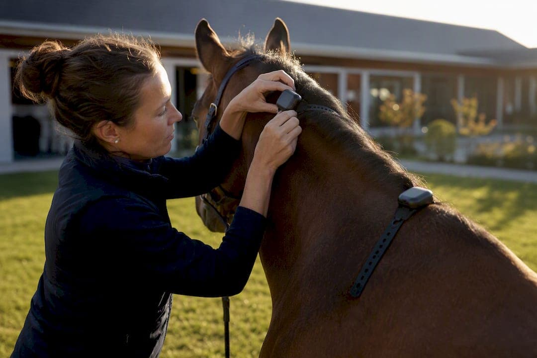
Horse movement analysis measures gait asymmetry and locomotor patterns using inertial measurement units (IMUs), markerless camera/AI systems, or lab-grade optical motion capture and force plates. IMUs remain the gold standard for high-precision, repeatable data in controlled settings, while markerless AI trades a little precision for field portability and remote monitoring. Neither replaces a clinical exam.
TL;DR:
- Markerless AI systems are useful for remote horse gait monitoring, but they depend heavily on lighting, background, and camera angles for accuracy.
- Inertial measurement units provide lab-quality data in the field and can monitor subtle asymmetries with minimal setup costs.
- Objective gait measures like HDmin and PDmin are most reliable when averaged over multiple strides and compared against the horse's own baseline.
- Consistent testing protocols, including fixed surface, camera setup, and metadata recording, are essential to minimize data noise and artifacts.
- Combining subjective clinical assessment with numerical gait data yields the most accurate diagnosis, especially when tracking trends over time.
Table of Contents
- What is horse movement analysis, and which technology fits your case?
- Reading the numbers: HDmin, PDmin, and what they actually tell you
- Building a repeatable data collection protocol
- Where the data goes wrong, and how to catch it
- Why an experienced eye still matters more than any algorithm
- Turning a single reading into a clinical decision
- Is there a standard protocol for measuring horse gait?
- Does conformation or discipline change what "normal" looks like?
- What's next for horse movement analysis?
- A practitioner's take on making objective data useful
- How EquiBETS turns gait data into a working clinical record
- Sources
What is horse movement analysis, and which technology fits your case?
Equine biomechanics research and everyday lameness work up now lean on three technology tiers, and each answers a different clinical question.
Optical motion capture (OMC) and force plates sit at the top of the accuracy ladder. They record three-dimensional joint kinematics and ground reaction forces with sub-millimetre precision, but they need a fixed lab, reflective markers, and a horse willing to trot over an embedded plate on cue. Few practices outside university hospitals run one.
IMUs (typically attached at the poll, withers, sacrum and sometimes the pelvis) capture vertical displacement and acceleration at 100 Hz or higher. A narrative review of inertial sensor systems found they can monitor and quantify locomotion with accuracy that matches or exceeds optical motion capture and force plate methods in many clinical tasks, at a fraction of the setup cost.
Markerless camera and AI systems use a smartphone or fixed camera plus a pose estimation algorithm to track anatomical landmarks (eye, withers, croup, hooves) frame by frame. No sensors, no markers, no wired connections. That convenience comes with a trade off: accuracy depends heavily on lighting, background clutter, and camera angle.
Choosing between them comes down to the question you're asking, not just budget:
- Lab research, kinetic validation, or biomechanics publication: OMC and force plates remain the reference standard.
- Routine lameness work-up, repeat monitoring, or a mobile practice: IMUs offer clinical-grade repeatability without a lab.
- Field screening, owner-submitted video, or remote check-ins between vet visits: markerless AI lowers the barrier to frequent capture.
- Rider-horse interaction studies: IMUs plus rider-side sensors can quantify how rider symmetry and weight distribution mechanically influence gait.
Reading the numbers: HDmin, PDmin, and what they actually tell you
Most lameness reports lean on a small cluster of vertical displacement metrics, and misreading them is one of the more common errors in interpreting objective data.
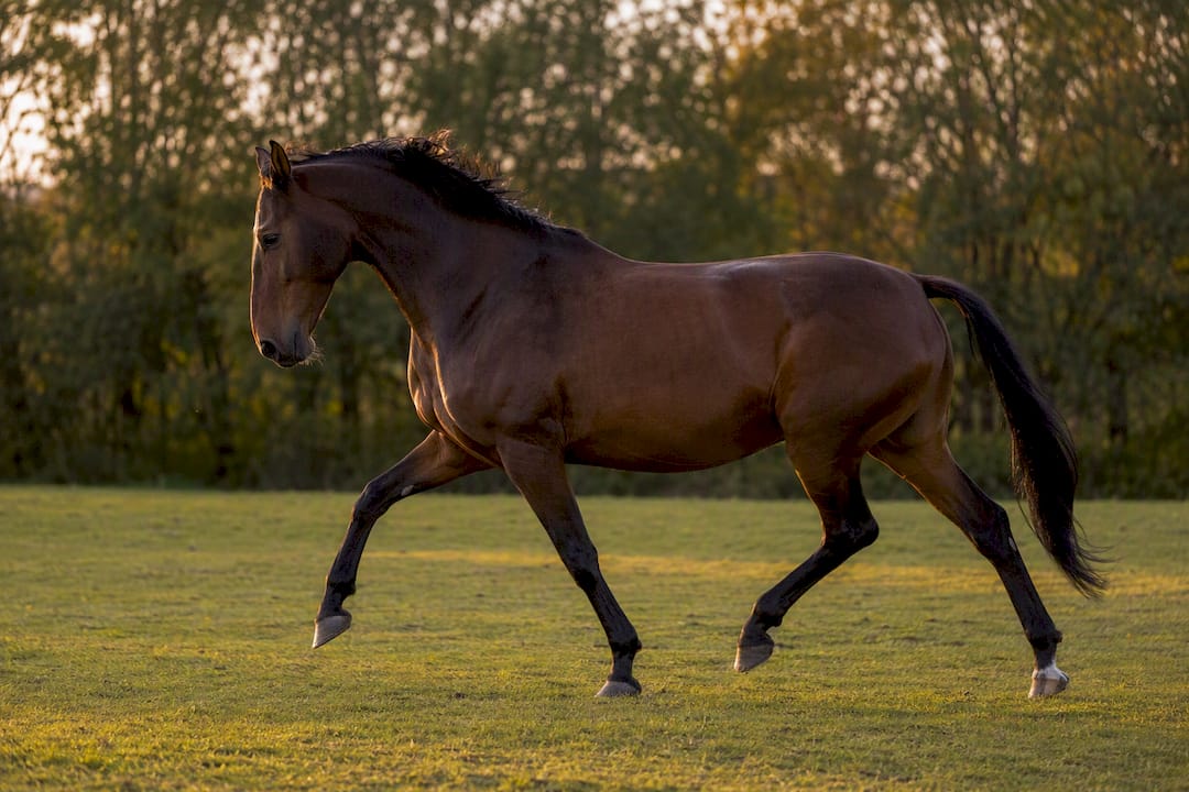
HDmin and HDmax describe the minimum and maximum height of the head during a stride cycle. In a forelimb lameness, the horse raises its head less when weight-bearing on the sound limb and lowers it more on landing of the sore one, so the difference between left and right minima (often written as Mindiff or HDmin diff) becomes the asymmetry signal clinicians chase.
PDmin and PDmax work the same way for the pelvis, tracking hindlimb push-off asymmetry via the tuber sacrale.
Maxdiff and Mindiff capture the absolute difference between left and right strides at the maximum and minimum points of the displacement curve. Range of motion (ROM), sometimes split into H-ROM and P-ROM, describes total vertical travel and helps flag reduced movement from stiffness or pain rather than pure asymmetry.
A validated markerless algorithm reported stride-level mean absolute errors of around 4.3 mm for Maxdiff and Mindiff, with frame-level keypoint accuracy ranging from 2.9 mm at the eye to 11.8 mm at the withers, a landmark that moves more with skin and coat.
Pro Tip: Don't treat a single trial's numbers as gospel. Some reviews reference thresholds like 12 mm for HDmin and 6 mm for PDmin, but these come from specific study populations and aren't universally agreed clinical cut-offs — always weigh a value against that individual horse's own baseline.
- Stride-level data shows trial-to-trial variability that trial-level averages can mask.
- Aggregate across multiple strides (ideally 25 or more) before drawing conclusions.
- Compare against the horse's own prior recordings, not a generic population figure.
Building a repeatable data collection protocol
Noise in your dataset usually traces back to the setup, not the technology. A tighter protocol fixes more problems than a better algorithm ever will.
- Choose a consistent surface. Concrete or firm, level asphalt gives the most reliable forelimb readings; soft or uneven arena surfaces increase variability, particularly for pelvic measures.
- Run straight-line trot-ups first. A validated field protocol calls for three straight passes of at least 30 metres each, trotted in hand at a consistent, moderate pace.
- Add lunging only when needed, using roughly 45 seconds per direction on a 15 to 20 metre circle, and interpret results with the understanding that circling itself introduces kinematic changes.
- Fix camera height and distance for markerless capture, roughly hip height, three to five metres from the trot line, with the horse crossing the frame perpendicular to the lens.
- Calibrate vertical scale using a known reference, such as the horse's withers height, before recording.
- Attach IMUs to standard landmarks (poll, withers, tuber sacrale) and confirm firm contact, since displacement or slippage during the trot is a major source of error.
- Log metadata every session: surface type, tack, handler, sedation status, and sensor sampling rate, typically 100 Hz or higher for IMUs.
- Record native frame rate for cameras, ideally 50 to 100 Hz, to capture clean vertical displacement signals rather than interpolated approximations.
Pro Tip: Keep the handler and route identical across repeat sessions. A different handler running at a slightly faster pace, or a trot line with a subtle camber, can shift Mindiff values enough to look like a clinical change when it's really a protocol artefact.
Where the data goes wrong, and how to catch it
Objective gait tools fail quietly, and the failures rarely look dramatic on the printout. A few conditions do most of the damage.
Groundline estimation is one of the more sensitive steps in any markerless pipeline. Inconsistent lighting, cluttered backgrounds, or a hoof landing in shadow can shift the algorithm's vertical displacement signal without any obvious visual sign to the clinician reviewing the output.
Wireless dropouts affect IMU trials less often but more severely. A missed data packet mid-stride can produce a spike that looks like a real asymmetry event.
Lungeing changes the horse's biomechanics on its own, even in a sound animal, because the circle itself loads the inside and outside limbs differently. Sedation dampens the horse's normal compensatory movement, which can flatten or exaggerate asymmetry depending on dose and drug.
- Displaced or loose sensors introduce artefacts that mimic clinical asymmetry.
- Soft or sloped surfaces degrade pelvic measures more than forelimb ones.
- A single trial, on its own, tells you far less than three repeat trials averaged together.
Objective gait analysis aids clinicians but does not replace subjective clinical assessment. Camera systems in particular require more trot-up repetitions and remain sensitive to environmental conditions that a trained eye would simply account for on the day.
Correlate any flagged asymmetry with a hands-on exam, flexion tests, and, where indicated, diagnostic analgesia before treating a number on a screen as a diagnosis.
Why an experienced eye still matters more than any algorithm
Traditional observational gait assessment, the trained eye watching a horse trot up, has one enduring advantage: context. A vet notices the ear pinning, the shortened stride length under saddle, the subtle head tilt that no vertical displacement sensor is built to capture. Decades of veterinary training went into building that pattern recognition, and it still catches things no camera currently tracks.
Its weakness is equally well documented. Human observers disagree with each other on subtle asymmetries, and fatigue, lighting, and observer bias all creep in over a long clinic day. Objective tools solve exactly that problem: consistency. An IMU or markerless system returns the same reading regardless of who is watching or how many horses came before it that morning.
The two approaches aren't really competing for the same job. Observational assessment excels at gestalt, catching the horse that "just doesn't look right" even when no single metric crosses a threshold. Objective measurement excels at quantification, turning a vague impression into a number you can track across weeks or compare against a treatment response.
The strongest work up combines both, in that order. Watch the horse first, form a clinical impression, then use IMU or markerless data to quantify what you saw, track it over time, and communicate it clearly to an owner or referring vet. Skipping the clinical look and jumping straight to sensor output risks over-interpreting noise as pathology, particularly in horses with mild or inconsistent gait changes.
Turning a single reading into a clinical decision
A single trot-up, however precisely measured, is a snapshot. The real value of horse gait evaluation comes from what you do with that number afterward.
Start by anchoring every reading against the horse's own history rather than a population average. Objective measures work best folded into longitudinal records that reveal small changes over time, rather than treated as a one-off pass or fail threshold.
Cross-check any flagged asymmetry against the physical exam before acting on it. A Mindiff spike on the left fore means little without a corresponding finding on palpation, flexion, or hoof testers. Where the picture is ambiguous, diagnostic analgesia (nerve or joint blocks) remains the tie-breaker between "measured asymmetry" and "clinically significant lameness."
Document context alongside every number. Surface, tack, sedation, handler, and even the horse's mood on the day all shift the reading, and a decision made without that metadata is a decision made on incomplete information.
Finally, set your own threshold for action based on trend, not a single crossing of a published cut-off. A horse sitting at 8 mm HDmin diff for three months, then jumping to 14 mm, tells a clearer clinical story than one reading of 14 mm in isolation. Machine learning classification built on IMU data has reportedly reached accuracy above 97% in controlled studies, but that figure assumes controlled placement and calibration, conditions you have to actively recreate in your own practice.
Is there a standard protocol for measuring horse gait?
Not a universal one, and that's a genuine gap in the field. Different labs and manufacturers still report Maxdiff, Mindiff, and ROM using slightly different stride-averaging windows, sampling rates, and sensor placements, which makes cross-study comparison harder than it should be.
Some convergence is happening. Validation papers increasingly report the same core metric set (HDmin/PDmin, Maxdiff/Mindiff, ROM) with documented measurement error, which lets clinicians at least judge how much confidence a given number deserves. The markerless field validation study recommends a specific minimal protocol, three straight trot passes of at least 30 metres plus two 45 second lunges per direction when circling is required, as a step toward comparable data across sites.
Sampling rate is another area edging toward agreement. Most current IMU protocols specify 100 Hz or higher, and camera-based systems increasingly target 50 to 100 Hz native capture rather than relying on interpolated frame rates.
What's still missing is agreement on clinical thresholds. A 12 mm HDmin or 6 mm PDmin cut-off appears in some published studies, but those numbers came from specific populations under specific conditions, and applying them blindly to a different breed, discipline, or surface is a documented source of misclassification. Until the field settles on population-adjusted or individually-baselined thresholds, the safest approach is treating any single published cut-off as a reference point, not a rule.
Does conformation or discipline change what "normal" looks like?
Yes, and this is one of the more underappreciated sources of false positives in gait interpretation. A horse's conformation sets its baseline movement pattern before any pathology enters the picture.
A horse with a naturally croup-high build or a longer back will show different pelvic displacement patterns than a compact, level-topped horse, even when both are perfectly sound. Limb length, angle of the pastern, and even head-neck carriage under saddle all shift the vertical displacement curves that HDmin, PDmin, and ROM are built from.
Discipline compounds this further. A dressage horse ridden in consistent collection develops a different stride pattern and head carriage than a racehorse or an endurance horse covering ground at a working trot. Reining horses, gaited breeds, and driving horses each bring their own baseline quirks that a generic population threshold simply won't capture accurately.
This is exactly why individual baselines matter more than population norms in day-to-day practice. A number that looks abnormal against a generic reference range might be entirely normal for that specific horse's conformation and job. Practitioners working across disciplines should expect to calibrate their interpretation, not just their equipment, to the horse and the sport in front of them.
What's next for horse movement analysis?
The direction of travel is toward more data, collected more often, with less specialist intervention required to get it.
Markerless AI is improving fastest. Comparative work already shows AI-based systems detecting more asymmetries than IMU setups in some conditions, with the strongest agreement on forelimb measures over hard, straight trot-ups and weaker agreement on pelvic measures over soft surfaces, a gap that better pose-estimation models should continue closing.
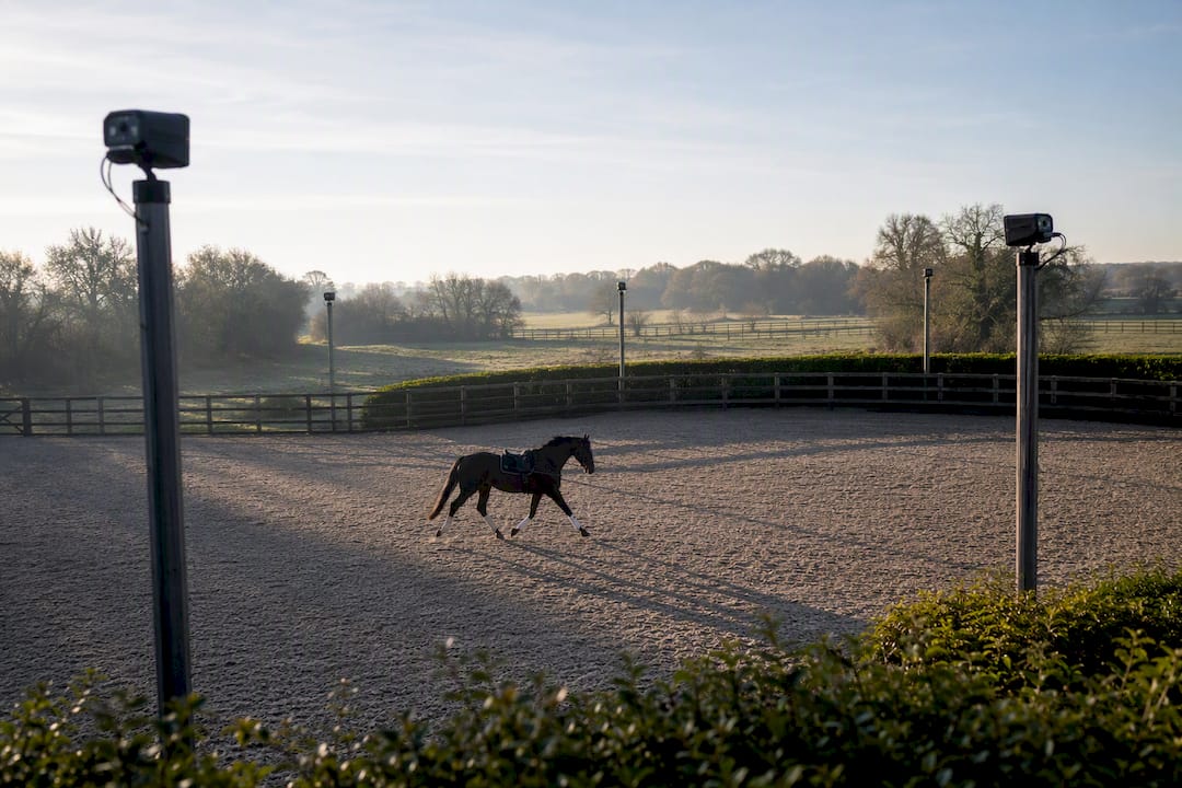
Owner-submitted video is quietly becoming a monitoring tool in its own right. Lowering the barrier to frequent capture lets training yards flag developing asymmetries earlier, between scheduled vet visits rather than only at them.
Expect wearable sensors to shrink further, sample longer without recharging, and integrate more directly with mobile apps that log metadata automatically rather than relying on a handler's memory. Multi-camera and depth-sensing setups are also starting to appear in research settings, aiming to close the gap between markerless field convenience and OMC-grade three-dimensional accuracy.
The bigger shift is structural rather than technological: moving gait data out of one-off PDF reports and into longitudinal records that live alongside a horse's training load, farriery, and veterinary history, which is where the next real gains in early detection will likely come from.
A practitioner's take on making objective data useful
The best use of a gait sensor isn't catching the dramatic lameness. It's the horse whose Mindiff crept up 3 mm a month for four months before anyone booked a vet visit. That trend, not a single trot-up, is what changed the treatment plan.
Build routine gait checks into your existing schedule, not as a separate appointment, and someone on the team needs to own the recording protocol so numbers stay comparable session to session. Objective measures earn their place by sharpening what you already do on exam, not standing in for it.
— isaac
How EquiBETS turns gait data into a working clinical record
A single trot-up video or IMU export is only useful if someone can find it again three months later, next to the horse's farriery notes and training log. That's the actual bottleneck for most yards, not the measurement itself.

Equibets is built around that gap. Its AI-powered movement analytics sit inside the same workspace as the horse's daily records, so a gait check taken this morning links directly to feed changes, workload, and prior veterinary notes rather than living in a separate app or a folder of loose video files. Offline capture means a yard can record a trot-up in a paddock with no signal and have it sync the moment the phone reconnects.
Key features relevant to a movement analysis workflow include:
- AI movement analytics linked to each horse's ongoing record, not a standalone report
- Offline mobile capture for yards without reliable arena connectivity
- Owner portals that share trend reports without a phone call
- Stable management tools that connect gait data to handover notes for vets and staff
- Nutrition and feed tracking that helps contextualise movement changes against diet and workload
The platform runs on a single subscription plan covering every module, with a free trial available before you commit. Import your existing horse records, run a trial period across your yard's actual caseload, and see whether longitudinal tracking changes how early you catch a developing issue. Check current plan details or start with the platform overview to see how it fits your workflow.
Sources
For deeper reading beyond this primer: the RVC Equine gait analysis service page covers clinical application; the inertial sensor narrative review details IMU validation; the markerless field reliability study sets out protocol and error rates; and the JAVMA review of objective gait methods offers clinical integration guidance.
- Gait Analysis - RVC Equine
- Narrative review on inertial sensors and applicability in equine gait analysis
- Reliability, agreement and variability of a markerless computer vision algorithm for equine gait analysis under field conditions
- Objective gait analysis: IMU vs markerless AI comparative study
- Objective gait analysis methods for assessing movement asymmetry in the equine patient