
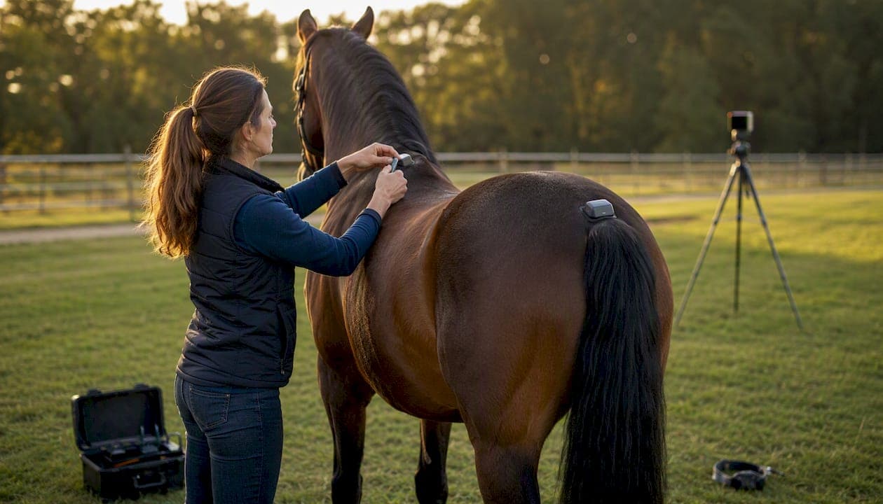
Equine gait analysis is the objective, millimeter-resolution measurement of a horse's movement to detect, quantify, and monitor locomotor asymmetry. The clinical verdict: use it as a complementary, evidence-based tool alongside the physical exam, not as a replacement for it. Two authoritative reference points anchor this guide:
- EGAS (Equine Gait Analysis Society): the primary professional body for training, standards, and certification in objective gait assessment
- EquiBETS: a clinic-ready management platform that integrates AI-assisted movement analysis, owner portals, and offline mobile recording into a single workflow
Key Takeaways
Objective equine gait analysis, used serially with consistent protocols, gives clinicians millimeter-resolution asymmetry data that visual examination cannot reliably detect.
| Point | Details |
|---|---|
| Accuracy below visual threshold | Validated systems report 3–7 mm precision, detecting asymmetry the human eye misses. |
| Serial measures over snapshots | Single-session data is a starting point; pre/post-intervention comparisons deliver the most clinical value. |
| Hindlimb data needs more caution | Pelvic measures show the lowest agreement between systems; confirm findings on hard, straight surfaces. |
| Protocol consistency is non-negotiable | Surface, sensor placement, and camera angle must be identical across sessions for meaningful comparison. |
| Equibets integrates the workflow | Equibets connects AI movement analysis, owner portals, and stable records in one platform for clinic-ready adoption. |
Table of Contents
- What is equine gait analysis and how do the main systems work?
- What does the evidence say about accuracy and repeatability?
- How do clinicians use gait data, and where does it fall short?
- What does a reliable recording protocol look like?
- How do you interpret core asymmetry parameters?
- How do you build gait analysis into clinic workflow?
- Where can clinicians get trained and what should they read first?
- What does the strongest evidence actually support?
- What actually changes when you adopt objective gait analysis
- Equibets supports the full clinic workflow, from pilot to daily use
- Sources
What is equine gait analysis and how do the main systems work?
Two technology families dominate objective horse movement assessment: inertial measurement units (IMUs) and camera-based computer vision. They capture different data, suit different environments, and carry different trade-offs.
IMU-based systems
IMUs measure linear acceleration and angular velocity in three axes. Sensors attach to the head, withers, and pelvis, producing the core output parameters clinicians rely on: head nod (MinDiffhead, MaxDiffhead), hip hike (MinDiffpelvis, MaxDiffpelvis), and symmetry indices. The JAVMA technical tutorial on objective gait analysis confirms IMUs offer good repeatability and field robustness, though precise sensor placement is non-negotiable and takes time to do correctly.
A review of inertial sensor technologies in equine gait analysis concludes these systems are practical for field monitoring and have supported gait-classification models with high reported accuracy in research settings. That combination of portability and precision makes IMUs the workhorse of clinic-based lameness quantification.
Camera-based and computer-vision systems
Marker-less AI systems extract body-landmark positions from standard video, with no sensor attachment required. The horse trots past a fixed or handheld camera, and the algorithm tracks key anatomical points frame by frame. Setup is faster than IMU placement, and the format allows owner-recorded video uploads for remote analysis, a genuine advantage for monitoring horses between clinic visits.
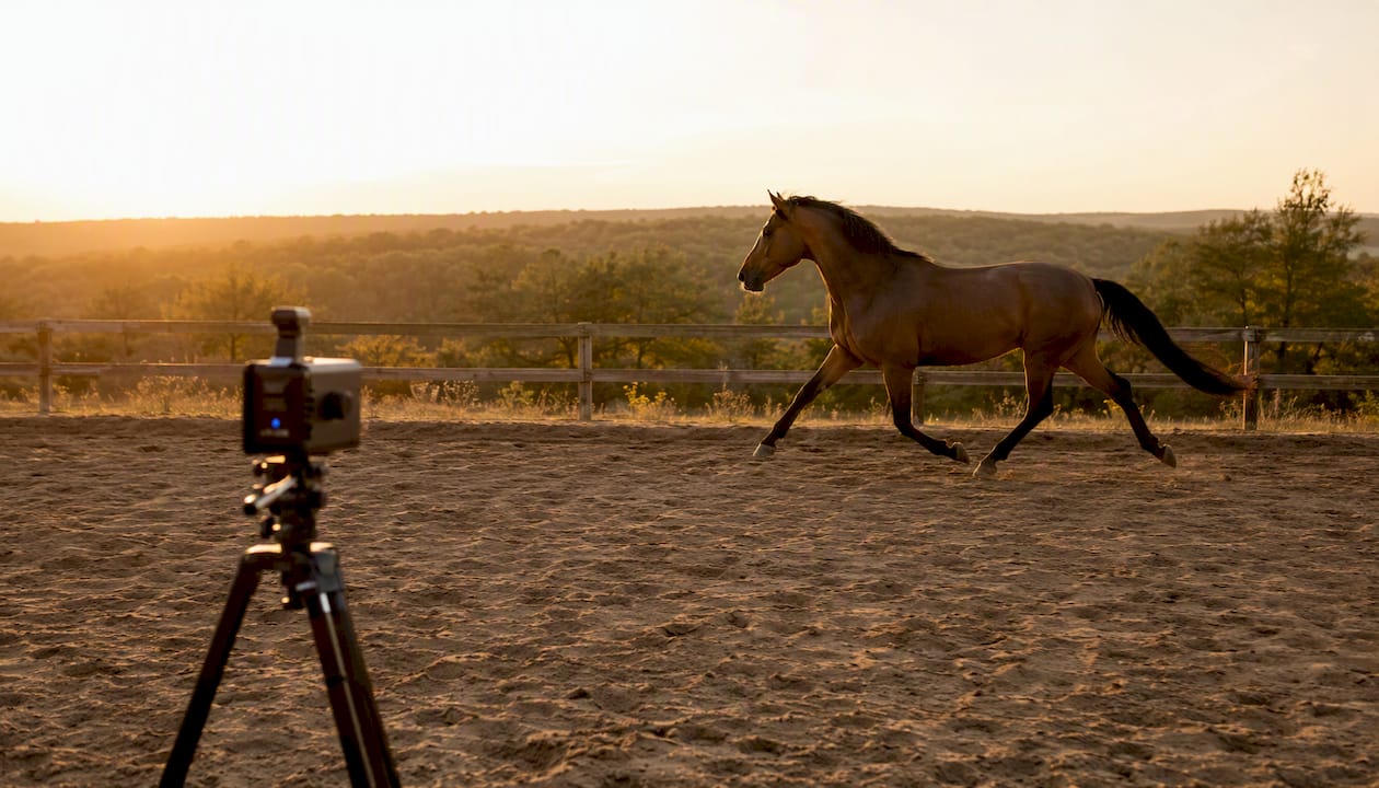
The trade-off is environmental sensitivity. Lighting quality, surface color, camera angle, and background clutter all affect tracking accuracy. A comparative study of IMU and AI marker-less systems found AI-based systems detected more asymmetries than IMUs and visual exam combined, with the strongest agreement on straight, hard-surface trot-ups and the weakest agreement for pelvic and hindlimb measures.
Practical trade-offs at a glance
- Setup time: IMUs require 5–10 minutes of careful sensor placement; camera systems are ready in under two minutes
- Cost: IMU kits typically have higher upfront hardware costs; camera systems may leverage existing devices with software subscriptions
- Environmental sensitivity: IMUs perform robustly across various field conditions; camera systems are more sensitive to lighting and environmental factors
- Remote use: Camera systems allow owner-submitted video uploads for remote analysis; IMUs generally require physical sensor attachment at a clinic or yard
- Hindlimb accuracy: Both IMU and camera systems show reduced reliability for pelvic measures compared to head metrics; IMU sensor placement precision is important to minimize error
Pro Tip: When a client wants to monitor a horse between appointments, camera-based video upload is a practical option. Brief the owner on lighting (outdoors, overcast preferred), surface (hard, level), and framing (full body in frame, side view) before they record. A poorly lit, shaky clip produces data no system can reliably interpret.
What does the evidence say about accuracy and repeatability?
The precision numbers matter here. Current objective systems report accuracy and repeatability in the range of 3–7 mm, which sits below the detection threshold of the human eye. That gap is clinically significant: a trained clinician watching a trot-up will miss asymmetries that a sensor records reliably.
What those millimeter values mean in practice depends on context. A 6 mm head nod asymmetry in a horse with no history of lameness reads differently than the same value in a horse three weeks post-nerve block. The number is only as useful as the baseline it is compared against.
Key evidence points:
- Agreement between systems is highest on straight-line, hard-surface trot-ups for head metrics and lowest for pelvic measures on soft surfaces
- The JAVMA tutorial notes IMUs can be inaccurate with mild hindlimb lameness, particularly when asymmetry is subtle and sensor placement is imperfect
- Thresholds for clinical relevance remain debated in the literature; no single cutoff value applies across breeds, disciplines, and individual horses
- Serial measurements and pre/post-intervention comparisons (flexion tests, diagnostic analgesia) provide far more interpretive power than any single snapshot
The practical implication: treat a single session's output as a starting point, not a verdict. The JAVMA tutorial and the IMU review both emphasize longitudinal monitoring as the highest-value application of these tools.
How do clinicians use gait data, and where does it fall short?
Clinical applications
Objective gait data earns its place in four main scenarios:
- Lameness detection and localization support: quantifying asymmetry that is subclinical on visual exam, or confirming the primary limb when compensatory patterns complicate the picture
- Flexion test and diagnostic analgesia quantification: measuring the magnitude of change before and after a block, giving a number to what was previously a subjective "improved" or "worse"
- Rehabilitation and training monitoring: tracking recovery trajectory over weeks or months, with data points that owners and referring vets can follow
- Horse-rider interaction assessment: emerging application using IMUs to assess how rider position and movement affect the horse's symmetry metrics
The Royal Veterinary College's gait analysis service illustrates what integration looks like at scale: wireless inertial sensors produce rapid summary reports that feed directly into specialist decision-making and pre/post-intervention comparisons.
Known limitations
- Mild hindlimb lameness is the hardest to detect objectively; sensitivity drops when asymmetry is subtle, and sensor placement errors compound the problem
- Lungeing introduces kinematic adaptations that can render head and pelvic movements asymmetrical in otherwise sound horses; circle data requires a different interpretive frame than straight-line data
- Surface and environmental sensitivity: soft or uneven ground increases measurement variability, particularly for pelvic metrics
- Sensor displacement during the trot-up, even a few millimeters of shift, can produce artifacts that look like genuine asymmetry
- Alpha-2 agonists can mask forelimb lameness; standard tranquilizers generally do not strongly affect gait metrics, but drug use should be documented and factored into interpretation
Always pair objective metrics with the clinical exam and, where indicated, imaging. A number that conflicts with the physical exam is a prompt for further investigation, not a tie-breaker.
Pro Tip: When you need to lunge a horse for clinical reasons, record straight-line trot-ups first and treat them as your reference. Lunging data is useful for detecting directional differences, but compare it only to other lunging data from the same horse, not to straight-line norms. Mixing the two in a single interpretation is one of the most common errors in clinical gait reporting.
What does a reliable recording protocol look like?
Consistency is the single biggest driver of data quality. Small changes in surface, sensor position, or camera angle create measurement errors that are large relative to the millimeter-scale signals being assessed.
Pre-test checklist
- Select a hard, level surface of at least 25–30 meters straight run; avoid deep sand or wet grass
- Check lighting: outdoors in diffuse daylight is ideal; avoid direct sun creating sharp shadows across the horse's topline
- Confirm battery charge and wireless connection for all sensors before the horse enters the area
- Remove or secure any loose equipment on the horse that could displace sensors during trot
- Brief the handler on pace (working trot, consistent speed) and straight line
Sensor placement (IMUs)
- Head sensor: attach at the midline of the frontal bone or poll, secured firmly with a purpose-made headband or adhesive pad
- Withers sensor: midline over the dorsal spinous processes of T3–T5, secured with a surcingle or adhesive
- Pelvis sensor: midline over the sacrum, secured with a tail bandage or adhesive pad; confirm it is level and not tilted laterally
- Run a brief calibration pulse (per vendor protocol) and verify signal quality before starting trot-ups
Camera setup
- Position the camera perpendicular to the trot-up lane, at mid-body height (approximately wither level)
- Distance: 8–12 meters from the horse's path, capturing the full body in frame throughout the pass
- Use a tripod; handheld footage introduces motion artifact
- Set frame rate to at least 60 fps; 120 fps is preferable for detailed stride analysis
Trot-up protocol
- Allow 3–5 minutes of walk warm-up before recording
- Record a minimum of 3–5 straight-line passes in each direction; most validated systems require at least 3 clean passes for reliable averaging
- If flexion tests are planned, record baseline passes first, apply flexion, then record immediately after release (within 60–90 seconds)
- For diagnostic analgesia: record pre-block baseline, wait the appropriate onset time per block site, then record post-block passes using the identical protocol
Immediate data quality checks
- Review signal traces for dropped packets, flat lines, or excessive noise before the horse leaves the area
- Confirm sensor orientation flags (most systems alert to tilted or displaced sensors)
- If a pass shows a sharp artifact mid-stride, discard it and record a replacement
Pro Tip: Always photograph sensor placement before the first trot-up. If you need to repeat the session a week later, that photo is the fastest way to replicate exact positioning. Positional consistency between sessions matters more than absolute placement precision.
How do you interpret core asymmetry parameters?
Head nod and hip hike are mechanistically linked to fore- and hindlimb lameness and are the two primary parameters objective systems quantify. Understanding what each metric represents prevents the most common interpretation errors.
Core parameters and what they indicate
| Parameter | Mechanical meaning | Common pitfall |
|---|---|---|
| MinDiffhead | Head position at minimum height: elevated when the lame forelimb is in stance | Confusing left vs. right convention across software platforms |
| MaxDiffhead | Head position at maximum height: reduced push-off from the lame forelimb | Misreading compensatory head movement as primary lameness |
| MinDiffpelvis | Pelvis at minimum height: reduced on the lame hindlimb side | High variability on soft surfaces; confirm on hard ground |
| MaxDiffpelvis | Pelvis at maximum height: reduced push-off from the lame hindlimb | Sensor tilt artifacts mimic genuine asymmetry; check placement photo |
| Symmetry index | Composite score of left/right difference | Does not distinguish primary from compensatory lameness without clinical context |
Using pre/post-intervention comparisons
The most defensible use of these numbers is change detection. A horse with a MinDiffhead of 12 mm pre-block that drops to 3 mm post-perineural analgesia of the palmar digital nerve has given you a clear localization signal. The magnitude of change matters more than the absolute value, and most validated systems flag changes above their own repeatability threshold as clinically meaningful.
Practical guidance:
- A change smaller than the system's reported repeatability range (typically 3–7 mm) should be treated as noise, not response
- When numbers conflict with the visual exam, recheck sensor placement and surface consistency before concluding the data is wrong
- Forelimb lameness cases tend to produce cleaner head metric data; hindlimb cases require more caution and more passes
For hindlimb cases specifically, the comparative study found pelvic measures show the lowest agreement between systems and between objective and visual assessment. That is not a reason to avoid measuring; it is a reason to weight serial measures and post-block comparisons more heavily than any single pelvic reading.
How do you build gait analysis into clinic workflow?
Adoption works best as a staged process. Rushing from equipment purchase to full clinic rollout without a pilot phase is the most common reason practices abandon these tools within six months.
Stepwise adoption checklist
- Pilot phase (weeks 1–4): Select 5–10 horses with known lameness diagnoses. Run parallel objective assessments alongside your standard exam. Compare outputs. Identify where the system adds information and where it does not.
- Staff training (weeks 2–6): Assign a primary operator for sensor placement and data acquisition. Use EGAS course materials and vendor-supplied tutorials. Budget 4–8 hours of hands-on training per operator before unsupervised use.
- Protocol standardization (week 4 onward): Write a one-page clinic protocol covering surface, sensor placement, camera setup, and trot-up count. Laminate it and post it at the recording area.
- Owner communication templates (week 6): Prepare a one-page visual summary that translates asymmetry scores into plain language. Owners respond better to trend graphs than raw millimeter values.
- Full rollout (week 8+): Integrate gait reports into the medical record as a standard exam component for lameness workups.
Staff roles
- Sensor operator: places and checks sensors, runs calibration, discards poor-quality passes
- Handler: maintains consistent trot pace and straight line
- Interpreting clinician: reviews output alongside clinical exam, documents findings in the record
Budget and timeline notes
Equipment costs vary by system type and vendor. IMU kits typically require a higher upfront hardware investment; camera-based systems often run on existing tablets or smartphones with a software subscription. Staff training to competence generally requires 4–8 hours of supervised practice per operator. A realistic pilot-to-full-adoption timeline is 8–12 weeks for a two-clinician practice.
Data management and privacy
- Store video files and sensor data in a HIPAA-aligned or equivalent secure system; equine patients are not covered by HIPAA directly, but client data attached to records is
- Obtain written owner consent before uploading video to cloud-based analysis platforms
- Confirm your vendor's data retention and export policies before signing a subscription; you need to be able to export raw data if you change systems
- Integrate gait reports as PDF or structured data exports into your practice management software; avoid siloed data that only lives inside the gait analysis platform
Where can clinicians get trained and what should they read first?
Training and certification
- EGAS: the primary professional training body for objective equine gait analysis; offers structured courses covering acquisition, interpretation, and clinical integration
- JAVMA continuing education: the 2026 technical tutorial is a peer-reviewed, practice-ready reference that covers both IMU and camera-based methods with clinical examples
- University CE programs: several veterinary schools offer short courses and webinars on equine biomechanics and objective lameness assessment; check with your state veterinary association for accredited options
- Vendor training: most IMU and camera system vendors provide onboarding tutorials and technical support; treat these as a starting point, not a substitute for peer-reviewed education
Core reading list
- PMC IMU review — systematic review of inertial sensor accuracy, field applicability, and research applications
What does the strongest evidence actually support?
The evidence base for objective horse gait analysis is solid on accuracy and repeatability, thinner on clinical decision thresholds, and still developing on long-term monitoring norms.
Evidence-backed takeaways:
- Objective systems detect asymmetry below the human visual threshold, consistently across multiple studies
- IMU repeatability is good on straight, hard-surface trot-ups; it degrades with soft surfaces, mild hindlimb lameness, and imprecise sensor placement
- Camera-based systems detect more asymmetries overall but show lower agreement with IMUs and visual exam for pelvic measures
- Forelimb lameness is more reliably quantified than hindlimb lameness across both technology families
- Serial measurements provide more clinical value than single-session snapshots
Research gaps to know:
- No universally agreed threshold for what magnitude of asymmetry is clinically meaningful across breeds and disciplines
- Long-term monitoring reference ranges (what is "normal" variation over a season for a sound horse?) are not yet established
- Breed- and discipline-specific norms are limited; most validation data comes from Warmbloods and Thoroughbreds in controlled trot-up conditions
The IMU review and the Pfau paper both point toward longitudinal monitoring as the next productive frontier. Clinicians who build baseline datasets now will be ahead of the curve when those norms arrive.
What actually changes when you adopt objective gait analysis
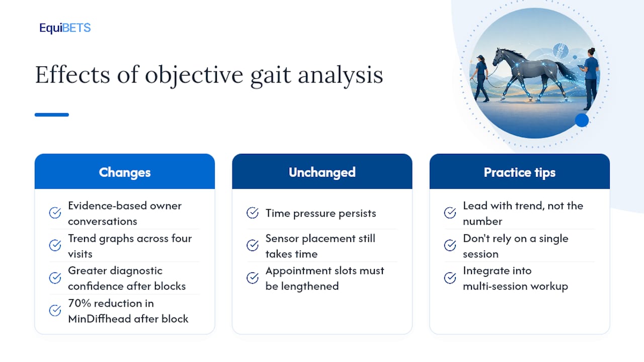
The first thing that changes is the owner conversation. When you can show a graph of asymmetry scores across four visits instead of saying "he looks a little better," the conversation shifts from subjective to evidence-based. Owners engage differently with numbers, especially when the trend line is moving in the right direction.
The second change is diagnostic confidence after nerve blocks. Before objective measurement, "improved" after a palmar digital block was a clinical judgment call. With a pre/post asymmetry score, you have a magnitude. A 70% reduction in MinDiffhead after a specific block is a different clinical statement than "he seemed to move more freely."
What does not change: the time pressure of a busy ambulatory day. Sensor placement still takes time, and that time has to be built into the appointment slot. Practices that try to add objective assessment without adjusting appointment length burn out their staff and cut corners on placement, which defeats the purpose.
Pro Tip: When presenting asymmetry scores to owners, lead with the trend, not the number. "His head asymmetry has dropped from 14 mm to 5 mm over three visits" lands better than "his MinDiffhead is 5 mm." The number means nothing to most owners; the direction means everything.
One caution worth stating plainly: a single session's data should never drive a major treatment decision on its own. The RVC's approach of integrating sensor data into a broader specialist workup reflects the right frame. Objective gait data is most powerful when it is one layer in a multi-session, multi-modality picture.
Equibets supports the full clinic workflow, from pilot to daily use
Objective gait analysis generates data. Managing that data, sharing it with owners, and keeping it connected to the horse's full health record is where most clinics hit friction. Equibets is built to remove that friction.

The Equibets platform combines AI-assisted movement analysis with offline mobile recording, owner portals, and stable records in one workspace. For a clinic running the adoption checklist above, that means gait reports, medical records, and owner communications live in the same system from day one of the pilot. No separate folders, no emailed PDFs that get lost, no manual re-entry.
The stable manager module handles record storage and team coordination, so the sensor operator, interpreting clinician, and yard manager all work from the same horse record. Owner portals deliver progress updates automatically, which cuts the time spent on post-appointment calls.
Equibets runs on a single monthly subscription covering all features and unlimited horses. A free trial is available before any paid commitment. Start your trial at Equibets and see how the workflow fits your clinic before you commit.
Sources
- PLOS ONE article on quantitative assessment of gait parameters
- Objective Assessment of Equine Locomotor Symmetry Using an Inertial Sensor System and Artificial Intelligence: A Comparative Study
- Quantitative assessment of gait parameters in horses: Useful for aiding clinical decision making? (Pfau)
- Gait Analysis - RVC Equine (Royal Veterinary College)